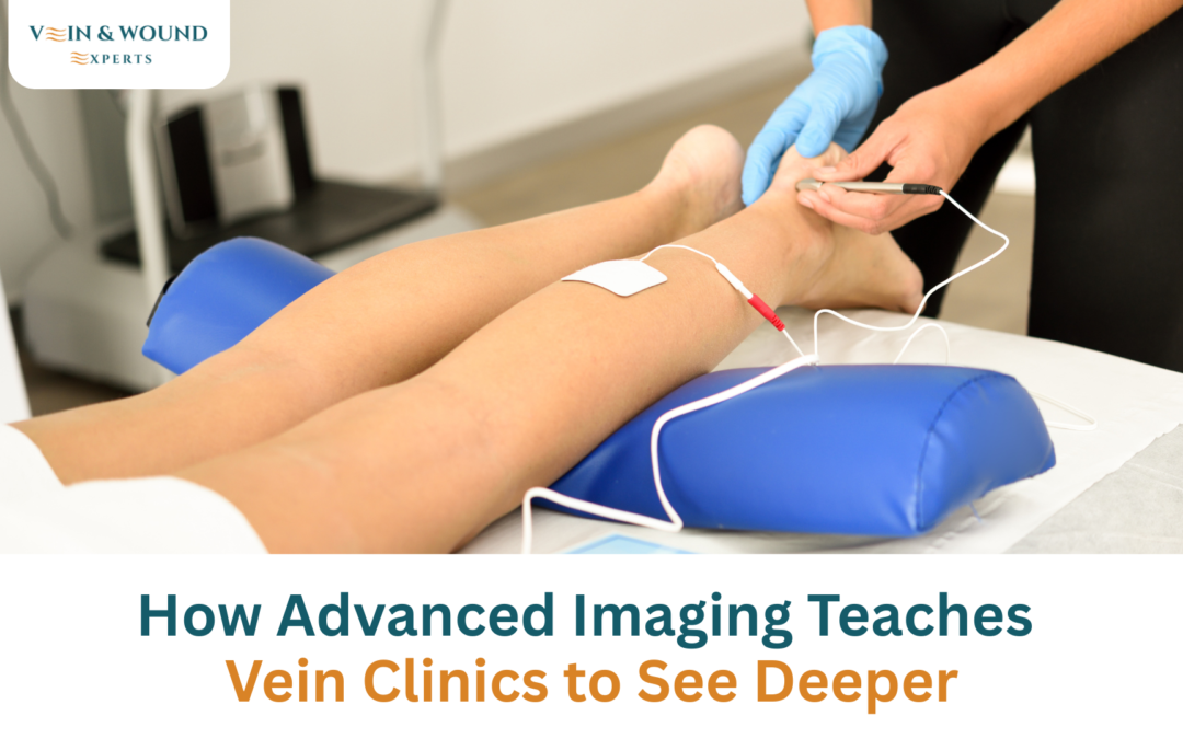
How Advanced Imaging Teaches Vein Clinics to See Deeper
Not every vein problem shows up as a bulging line on the leg. Some patients feel heaviness, swelling, aching, or skin discomfort long before anything obvious appears on the surface. That is where advanced imaging becomes important. It helps vein specialists see how blood is moving beneath the skin before choosing the right treatment.
👀 Why Visual Examination Alone Is Not Enough
A leg exam can reveal visible clues, but it cannot show the full picture of vein function.
Hidden Vein Disease
Some vein problems begin deeper inside the leg. The surface may look normal, but the valves inside the veins may not be working properly. When blood does not move upward as it should, pressure can build in the lower legs. Over time, this may lead to swelling, heaviness, visible vein changes, or skin problems. Without imaging, these hidden issues can be easy to miss.
Symptoms Without Visible Veins
A patient may have leg discomfort even when there are no large visible veins. Symptoms such as aching, fatigue, ankle swelling, cramping, restless legs, or pressure after standing can still point to a circulation problem. This is why a visual exam alone may not be enough to explain what the patient is feeling. The concern is not always what can be seen. Sometimes, the real problem is how the veins are functioning.
Importance of Accurate Diagnosis
Accurate diagnosis helps prevent guesswork. If treatment is based only on appearance, the main source of the problem may be left untreated. Advanced vein diagnostics help identify whether symptoms are coming from surface veins, deeper valve issues, or another circulation concern. This gives the specialist a clearer path before recommending care.
🔍 The Role of Duplex Ultrasound
Duplex ultrasound is one of the most important tools used in modern vein evaluation. It allows specialists to see the veins and assess how blood is moving through them.
Mapping Blood Flow
Vein ultrasound mapping helps create a clearer picture of the patient’s circulation. The test can show which veins are carrying blood properly and which ones may be under pressure. It also helps identify the direction of blood flow, which is important when checking for valve problems. This information helps the specialist understand the condition beyond what is visible during an exam.
Detecting Venous Reflux
Venous reflux happens when blood flows backward instead of moving upward toward the heart. This can occur when vein valves are weak or damaged. Venous reflux imaging helps detect this backward flow and shows where the problem is happening. Finding reflux matters because it can explain symptoms like heaviness, swelling, aching, and visible vein enlargement.
Identifying Underlying Causes
Two patients may have similar symptoms but different causes. One person may have small surface veins. Another may have deeper valve dysfunction. Another may have swelling linked to a more advanced circulation issue. Duplex ultrasound helps separate these possibilities so the treatment plan targets the actual source of the problem.
🧭 How Imaging Improves Treatment Outcomes
Better imaging can lead to better treatment decisions. It helps specialists plan carefully instead of treating every visible vein the same way.
Personalized Planning
A treatment plan should be based on the patient’s vein structure, symptoms, and blood flow pattern. Imaging helps identify which veins need attention and which ones are working normally. This allows the specialist to recommend treatment that fits the patient’s condition rather than using a one-size-fits-all approach. For some patients, care may involve compression support or monitoring. For others, a procedure may be appropriate.
Avoiding Unnecessary Procedures
Advanced imaging can also help avoid treatments that are not needed. If a visible vein is not the main source of symptoms, treating it alone may not provide the relief the patient expects. Imaging helps confirm whether a procedure is necessary and which vein should be treated first. This is especially important for patients who have recurring symptoms or have had previous vein treatment.
Monitoring Treatment Success
Imaging may also be used after treatment to check progress. Follow-up ultrasound can help confirm whether a treated vein has closed properly and whether blood is moving through healthier pathways. This gives both the patient and specialist more confidence in the results. It also helps guide any next steps if additional treatment is needed.
🏥 Advanced Diagnostics at Vein & Wound Experts
Vein & Wound Experts uses advanced diagnostic tools to look beyond surface symptoms. During evaluation, the team may use duplex ultrasound to check blood flow, map problem veins, detect reflux, and identify the underlying cause of symptoms. This helps create a treatment plan based on evidence rather than appearance alone.
For patients with swelling, leg heaviness, aching, visible veins, or skin changes, imaging can provide answers that a basic visual exam cannot. The goal is simple: find the real source of the problem before starting treatment, so care is more targeted, appropriate, and personalized to the patient’s vein health.

❓ Frequently Asked Questions
Why Is Ultrasound Used in Vein Treatment?
Ultrasound is used to see how blood moves through the veins and whether the valves are working properly. It helps specialists identify problem veins before recommending treatment.
Is Vein Mapping Painful?
Vein mapping is usually not painful. It is a noninvasive ultrasound exam that uses sound waves to evaluate blood flow and vein function.
How Long Does an Ultrasound Take?
The time can vary depending on the patient’s symptoms and how many areas need to be examined. Most vein ultrasound exams are completed during the diagnostic visit.
Can Imaging Detect Hidden Vein Problems?
Yes. Imaging can help detect problems that may not be visible on the surface, including valve weakness, backward blood flow, and deeper circulation issues.
Andy Sharifi
Position