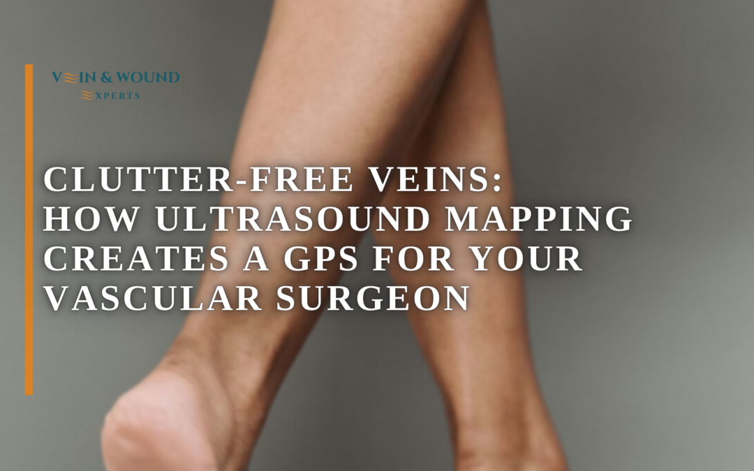
Clutter-Free Veins: How Ultrasound Mapping Guides Our Downey Surgeons 🩺🗺️
This technology allows doctors to study the veins beneath the skin in real time. By visualizing circulation patterns, specialists can locate damaged veins, track blood flow, and plan treatments with greater accuracy.
🔬 What Vein Ultrasound Mapping Actually Shows
Veins are part of a complex network responsible for returning blood from the body back to the heart. When certain veins weaken or valves stop functioning properly, blood may begin to pool instead of flowing smoothly.
Ultrasound imaging helps doctors observe this process. Using sound waves, the device creates live images of veins on a screen. Specialists can see how blood moves through each vessel and identify areas where circulation slows or reverses direction.
This technique is painless and non-invasive. A small handheld probe moves across the skin while the machine displays the internal vein structure and blood movement in real time.
🧭 Why Mapping Veins Is Important Before Treatment
Every person’s vascular system is unique. Without proper imaging, it would be difficult to determine which veins are functioning properly and which ones require medical attention.
Vein mapping acts like a navigation system for the vascular specialist. It helps pinpoint the exact location of problem areas so treatment can focus only on the veins that are not working correctly.
This detailed evaluation also helps prevent unnecessary procedures. By identifying the precise source of circulation problems, doctors can design targeted treatment plans that protect healthy veins while addressing damaged ones.
⚠️ Signs That May Lead to a Vein Evaluation
Many people seek vascular evaluation after noticing symptoms that suggest circulation problems. These symptoms often appear gradually and may worsen with time if ignored.
- 🦵 Persistent leg heaviness after standing or sitting for long periods
- 💧 Swelling around the ankles or lower legs
- 🌙 Nighttime leg discomfort or throbbing sensations
- 🕸️ Visible spider veins or bulging veins near the skin surface
- 🔥 Warm or aching areas along certain veins
These signs often prompt physicians to perform diagnostic imaging to better understand what is happening inside the vascular system.
🏥 How Specialists Use This Technology in Clinical Care
In specialized vascular clinics, ultrasound imaging plays a central role in evaluating circulation health. Experienced specialists use this diagnostic tool to trace blood flow, measure vein function, and identify where vein valves may be failing.
The imaging process also guides treatment planning. By studying the mapped veins beforehand, doctors can perform procedures with greater precision and efficiency.
This level of visualization allows specialists to focus only on the veins responsible for symptoms, improving outcomes and minimizing unnecessary intervention.
🌿 Clear Imaging Leads to Smarter Vascular Care
Accurate diagnosis is the foundation of effective vein treatment. With modern imaging techniques, specialists can observe circulation patterns that were once impossible to see without invasive procedures.
Vein ultrasound mapping provides the clarity needed to understand how blood moves through the legs, helping physicians identify damaged vessels and design precise treatment strategies.
For patients experiencing leg discomfort, swelling, or visible veins, advanced imaging technology offers a clearer path toward restoring healthy circulation and improving overall vascular wellness.

Andy Sharifi
Position